Overview
Measurement principle for γ-ray emitting nuclides
(germanium semiconductor detector)
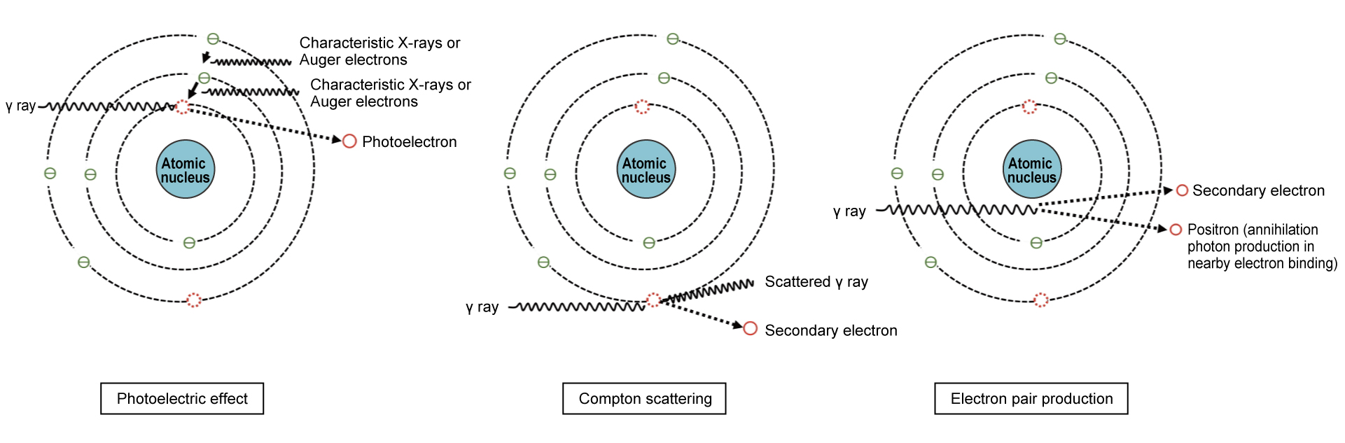
γ-ray emitting nuclides are measured indirectly by detecting the high-speed electrons (secondary electrons) that are produced as a result of the interaction between the incoming γ rays (photons) and the detector. γ rays entering the detector cause various interactions (photoelectric effect, compton scattering, electron pair production, etc.) to occur. Secondary electrons are produced, and the ionization effect of these electrons creates electron-hole pairs in the germanium crystal, which act as a detector by collecting these charges and converting them into electrical signals.
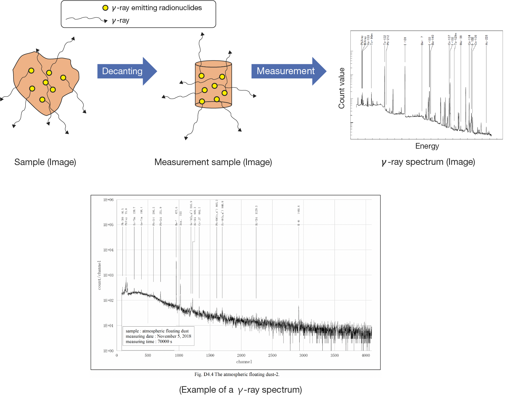
The observed energy spectrum (hereinafter referred to as the “spectrum”) detected by a γ-ray detector is primarily formed by single or multiple interactions occurring within the sensitive volume of the detector. However, it also includes contributions from other radiation—such as scattered γ-rays and X-rays—produced by interactions in surrounding materials, including shielding. When measuring a sample's γ-ray spectrum, the resulting spectrum is complex due to overlapping energy distributions produced by the interaction between the γ rays and the detector, as there are multiple γ rays of different energies present. However, only the photoelectric peak (total energy absorption peak) is analyzed. γ-ray spectrometry using a germanium semiconductor detector has excellent energy resolution and can simultaneously quantify multiple nuclides without requiring chemical separation. Of the seven main nuclides*1 in the ALPS-treated water,134Cs,137Cs,106Ru,60Co, and 125Sb have been measured using this method.
*1Tokyo Electric Power Company Holdings, Inc. Changes to the operational structure for the release of ALPS-treated water into the ocean and selection of nuclides for measurement and assessment [Summary]. 2023. https://www.nra.go.jp/data/0004208F97.pdf,(Accessed 2024-10-1)
Analysis flow
Seawater
← Precise analysis of134Cs, and 137Cs
Sample packing
Measurement
Addition of hydrochloric acid
Addition of phosphomolybdic acid
Filtration and separation
Sample packing and drying
Measurement
Main nuclides measured, γ-ray energy and emission fraction (table)
Nuclear data for nuclides with an emission fraction of 1% or more are listed.
Values for half-life, γ-ray energy, and emission rate are rounded to one decimal place.
| Nuclide name | Half-life | Half-life unit | γ-ray energy (keV) | Emission rate (%) |
|
60Co |
1925.3 |
Day |
1173.2 |
99.9 |
|
1332.5 |
100 |
|||
|
106Ru*2 |
371.8 |
Day |
511.9 |
20.4 |
|
621.9 |
9.9 |
|||
|
1050.4 |
1.6 |
|||
|
125Sb |
2.8 |
Year |
35.5 |
4.4 |
|
176.3 |
6.8 |
|||
|
380.5 |
1.5 |
|||
|
427.9 |
29.6 |
|||
|
463.4 |
10.5 |
|||
|
600.6 |
17.7 |
|||
|
606.7 |
5.0 |
|||
|
636.0 |
11.2 |
|||
|
671.4 |
1.8 |
|||
|
131I |
8.0 |
Day |
80.2 |
2.6 |
|
284.3 |
6.1 |
|||
|
364.5 |
81.5 |
|||
|
637.0 |
7.2 |
|||
|
134Cs |
2.1 |
Year |
475.4 |
1.5 |
|
563.2 |
8.3 |
|||
|
569.3 |
15.4 |
|||
|
604.7 |
97.6 |
|||
|
795.9 |
85.5 |
|||
|
802.0 |
8.7 |
|||
|
1168.0 |
1.8 |
|||
|
1365.2 |
3.0 |
|||
|
137Cs |
30.1 |
Year |
661.7 |
85.1 |
*2 For106, the data of 106Rh in radioactive equilibrium are provided.
Topics
Topics1
Energy calibration
Energy calibration determines the relationship between the observed peak center channel and γ-ray energy.
Correct calibration enables accurate γ-ray energy (E) determination, typically within 0.1 keV. Given the
excellent linearity between pulse height (P) and energy in germanium semiconductor detectors, a simple
linear equation (E=a+b・P) suffices for a practical energy calibration equation. For general radioactivity
analysis, adjust the amplifier gain, etc., to achieve a 4000-channel range for 0 to 2000 keV, setting a ≈
0 and b ≈ 0.5 keV/ch.
Accurate energy calibration is crucial for correct nuclide identification. However, consistent maintenance
is more important than frequent calibration. For normal operation, periodically check the position of a
main peak (e.g., 40K) in sample spectra. Calibrate if a deviation of more than 2 channels is observed.
Topic2
Self-absorption
In the case of volume samples, the phenomenon in which γ rays are attenuated by scattering or absorption in the sample medium is called self-absorption. It is a complex phenomenon that depends on the γ-ray energy, the type of sample medium (element composition, bulk density), the shape and thickness of the sample, and other geometric conditions, as well as the shape and size of the germanium crystal. The self-absorption of γ rays in volume samples can be as high as several tens of percent or more. Consequently, self-absorption correction is necessary when analyzing radioactivity in volume samples using γ-ray spectrometry.
Topic3
What are the main causes of BG spectra?
Background spectra (hereinafter referred to as “BG”) are mainly caused by the following factors:
1. Naturally occurring nuclides such as 40K, uranium-series isotopes (e.g., 214Pb and 214Bi), and thorium-series isotopes (e.g., 228Ac and 208Tl) are present in the structural materials of the measurement room (e.g., concrete). In addition, decay products of 222Rn (such as 214Pb and 214Bi) exist in the air within the room. γ rays from external sources can be significantly attenuated by thick lead shielding (5 to 15 cm).
2. Positron annihilation radiation (511 keV) is generated by cosmic rays and by electron-positron pair production from high-energy γ rays, such as those from 214Bi at 1764 keV and 2204 keV.
3. Ordinary lead shielding contains trace amounts of 210Pb (half-life: 22.3 years). Its decay product, 210Bi, emits β rays (1160 keV), which in turn produce bremsstrahlung X-rays. These X-rays contribute to the BG spectrum as a continuous distribution in the energy region below several thousand keV.
4. When γ rays induce the photoelectric effect on the inner surface of the lead shield, characteristic X-rays of lead (75.0, 72.8, and 84.9 keV) are detected. If these X-rays interfere with measurements below 100 keV, they can be suppressed by lining the inner surface of the lead shielding with another material—such as oxygen-free copper—about 1 mm thick.
Topic4
Cs collection efficiency of ammonium phosphomolybdate (AMP) with different grain sizes
When the same amount of AMP is added, smaller particle sizes result in higher Cs collection rates. This is considered to be due to the larger surface area per unit weight associated with smaller particles. However, care must be taken, as smaller particles sediment more slowly and are more likely to clog the filter paper.
Related radioactivity measurement series
No.13